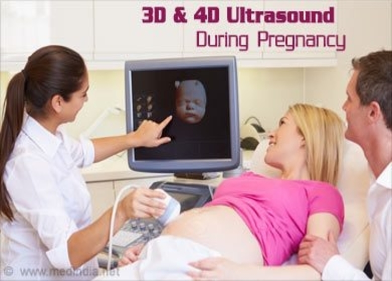
Know About 3D And 4D Ultrasound Scans
Seeing your unborn baby in its nearly actual form is always magical. Seeing this creates some connection with the parents who are eagerly waiting for a glimpse of their baby. This is made possible in a very realistic way by 3D and 4D Ultrasound scans.
What Are Ultrasound Scans In 3D And 4D?
Ultrasound scans like 3D and 4D are like your baby’s pictures in real time. Multiple baby images are taken in 2D in a 3D scan and then put together to create a 3D image effect. The pictures are taken in real time in a 4D scan, and at that point in time, you can see what your baby is doing inside you, like moving his legs and arms or even opening and closing his eyes. It’s more like a streaming live or a video. The time component is the fourth dimension added to the 3D scan and is therefore referred to as a 4D scan.
Why Do You Need 3D And 4D Scanning For Pregnancy?
Ultrasound scans are an important tool for monitoring a growing fetus ‘ internal organs and health. They help the gynecologist to determine whether or not the baby has any complications. This identification helps them to treat the baby as soon as possible. These scans help the doctor monitor the baby for any abnormalities like cleft lip, problems with the spinal cord and other birth defects. They also help the doctor to monitor the levels of amniotic fluid.
Difference Between 2D, 3D And 4D Scanning
2D Ultrasound Scan:
A decade ago, pregnant moms were able to see their babies before they were born using 2D scans. However, there were some limitations on 2D scans. One could only see the baby’s gray and hazy pictures. Such scans were only useful in viewing the internal organs as the waves of ultrasound used to create images of the organs of the baby and not their full form.
3D Ultrasound Scan:
Mothers – to – be are much luckier these days and can see how they look like their little – one before birth. 3D Ultrasound scans are much better than 2D scans, putting together the pieces of the scanned photos and creating a more real baby shape. You can see the face, legs, and arms of your baby.
4D Ultrasound Scan:
4D scans have improved technology and software further and help you see your baby (live) in real time. Now, the future parents can see their cutie’s feet move, yawn, kick, blinker and suck. It’s a delight to see what baby scanning technology has to offer. Although all of these scans are done for medical reasons, they help build a bond between the parents and the baby, and nothing can match the parents ‘ joy.
When 3D And 4D Scans Are Needed?
These ultrasonic scans are optional. They are not included in the prenatal tests. It’s perfectly fine if you don’t want any of these scans, and you can keep your doctor informed about the same. If she sees a medically relevant need or a probable complication, the doctor will advise you. Usually, it is done within 26 weeks to 30 weeks if you decide to have a scan. It’s best not to be taken earlier because under his / her skin the baby wouldn’t have fat. So, you may not get clear pictures of your baby.
How Are 3D And 4D Scanning Work?
A sonography scan is also called an ultrasound scan since high – frequency sound waves are used to create image slices for the infant. That’s how scans like that work:
- A transducer or a test is a gadget used to send ultrasound motions inside the belly. The transducer is first covered with a conductive gel that enables the waves to enter easily.
- The ultrasound signals, after experiencing a snag or structure, are reflected back, and the software captures them as pictures on the PC screen.
- The quality and time of the reflecting waves make pictures, and this data is caught as pictures or recordings.
- These slices are then collected to get a photo of the infant.
Results Of 3D And 4D Scans:
These scans are not standard prenatal test diagnostic procedures. They are merely indicative of possible birth defects or complications. Therefore, interpreting the results is up to the doctor. Further investigative tests will be advised if she/he notices any possible complications.
How Often Is The Scan Done?
Doctors regularly do a 2D scan to keep track of the progress of the baby. Knowing the vitalities of the baby, amniotic fluid levels, and the growth of the baby is usually a good test. It is not recommended to scan 3D and 4D as much as the 2D scans. Only to study the fetus closely and look for any birth defects are they done. Although no reliable evidence suggests that 3D and 4D scans might in any way harm your baby or mother, it is advisable to limit the scans and consult your physician before choosing private scans.
Advantages Of 3D And 4D Scans:
3D Scans Advantages:
- 3D scans better visualize fetal cardiac structures because it projects views that can not be accessed through 2D imagery.
- Helps to diagnose fetal facial defects like a cleft lip.
- It helps to diagnose musculoskeletal or neural defects in the fetus.
- Getting standard plane visualization takes less time.
- To diagnose common fetal anomalies, it is easy to study and does not require a very skilled or experienced person.
- For expert opinion and better diagnosis, the recorded volume data can be made available virtually.
4D Scans Advantages:
- Shorter time for screening and diagnosis of the fetal heart.
- Data obtained during the scan may also be sent to doctors residing in remote areas for expert examination.
- It can also be used for the purposes of training.
- Enhanced bonding with the baby by parents.
- Positive bonding with the baby as a result of seeing the baby in real time during pregnancy.
- After seeing the form and movement of the baby, families, and fathers are more supportive.
- The likelihood of more accurate identification of fetal anomalies in 4D scans is higher.
- It also shares the benefits of 3D ultrasound, such as:
- Evaluation of fetal growth and well-being
- Placental location and evaluation
- Examining the heartbeat of the fetus
Disadvantages Of 3D And 4D Scans:
3D Scans Disadvantages:
- In pregnancy, the only setback in 3D sonography is that it does not give us real-time or live streaming of the movement of the baby.
- High-speed computing software is required for 3D baby scanning.
4D Scans Disadvantages:
- Costly machinery.
- To operate the machinery, intensive training is required.
- The data acquired may be of lower quality in the presence of fetal movements.
- This, in turn, has an impact on all later viewing slices.
- If the fetal spine is not at the bottom of the scanned field, the view may be hindered by sound shades.
Although these scans are not part of the standard medical procedure, seeing your baby moving within you is always a pleasure. 3D and 4D scans are among parents ‘ most sought – after scans. Seeing your growing baby is magic. If during your pregnancy you want to perform these scans, consult your doctor and follow his advice.
Also Read: Sonogram nails – the new pregnancy trend


